Morphology

TCD Waveform Indices

Morphology provides physicians with advanced analysis of TCD waveforms to enable faster and easier assessment of patients with suspected neurological conditions, including stroke. Waveforms are collected using the NovaGuideTM 2 Intelligent Ultrasound with post-exam analysis available in NovaGuide View. Morphology features include Velocity Curvature Index, Velocity Asymmetry Index, Systolic Slope, and P2Ratio.

A labeled TCD waveform.
Cardiac Cycle Phase
Metric
Symbol
Unit
Definition
Systolic Upstroke
(Phase 1)
Systolic Slope*
-
cm/s2
(P1-V1) / (TP1-TV1)
First Systolic Peak
P1
cm/s
P1
Windkessel Notch
V2
cm/s
V2
Late Systole
(Phase 2)
Second Systolic Peak
P2
cm/s
P2
P2Ratio*
-
ratio
P2/P1
Diastole
(Phase 3)
Dicrotic Notch
V3
cm/s
V3
Third Peak
P3
cm/s
P3
End Diastolic Velocity
EDV (V1)
cm/s
V1
Full Cycle
Velocity Curvature Index*
VCI
-
Sum of instantaneouscurvatures of the beat canopy (upper 3/4 of the waveform, shown in purple)
*Morphology features

Velocity Curvature Index (VCI)

Definition: VCI is a measure of the overall curvature of a TCD waveform. A lower value of VCI represents a more blunted waveform.

Physiologic Relevance: VCI is an objective method to quantify changes in the TCD waveform that can occur due to various stroke pathologies.1

Bilateral Velocity Curvature Index (MVP-VCI)

Definition: Using the maximum velocity segments from each hemisphere, MVP-VCI looks at bilateral VCI values and reports the lower VCI number.

Physiologic Relevance: The extent to which the blood flow is disrupted and the resulting impact on the TCD waveform can be different on the left and right hemispheres of the brain. When bilateral measurements are available, MVP-VCI generally describes the waveform ipsilateral to the neurologic condition.1-3

Bilateral Velocity Asymmetry Index (MVP-VAI)

Definition: MVP-VAI is the ratio of mean velocities from a set of bilateral TCD waveforms. The maximum velocity segment from each side is used to minimize the effect of the angle of insonation.

Physiologic Relevance: MVP-VAI represents the level of blood flow velocity disparity between cerebral hemispheres. Quantifying the degree of hemispherical asymmetry in blood flow velocity could provide information on the severity and location of a neurological condition.4,5

Systolic Slope

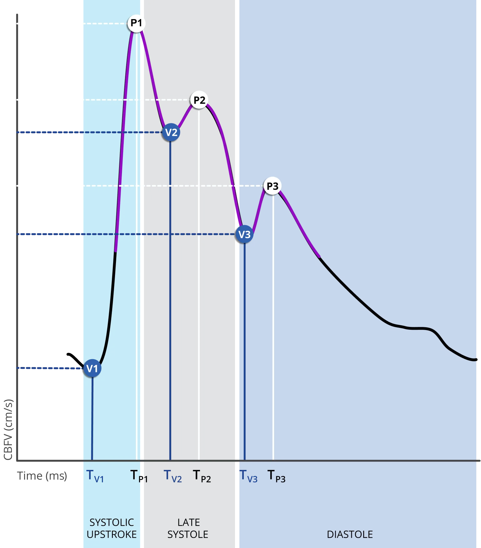
Definition: Systolic Slope is the average rate of change in velocity for the systolic upstroke phase of the cardiac cycle.

Physiologic Relevance: Systolic Slope has been reported to be inversely related to vascular resistance to blood flow. A low systolic slope could be indicative of obstruction to flow.6-8

P2Ratio

Definition: P2Ratio is the ratio of the second peak to the first peak of the TCD waveform.

Physiologic Relevance: P2Ratio is hypothesized to be related to the distal vasculature’s compliance. An elevated P2Ratio could indicate decreased compliance in the distal vasculature.9

Interested in learning more about Morphology?

Download the Morphology Interpretation Guide

DOWNLOAD THE GUIDE

References:

  1. Thorpe SG, Thibeault CM, Wilk SJ, et al. Velocity Curvature Index: A novel diagnostic biomarker for Large Vessel Occlusion. Translational Stroke Research. Published online 2018. doi:10.1007/s12975-018-0667-2
  2. Dorn AY, Thorpe SG, Jalaleddini K, Wilk SJ, Hamilton RB. Feasibility study in the combination of non-contrast computed tomography and transcranial Doppler for large vessel occlusion detection. Neurology and Neurosurgery. 2020;3(3). doi:10.15761/NNS.1000130
  3. Thibeault CM, Dorn AY, Thorpe SG, et al. A Case Study on the Utility of Transcranial Doppler Ultrasound in Acute Stroke Evaluation. Journal for Vascular Ultrasound. 2020;44(1):25-27. doi:10.1177/1544316719887842
  4. Zanette EM, Fieschi C, Bozzao L, et al. Comparison of Cerebral Angiography and Transcranial Doppler Sonography in Acute Stroke.; 1999. http://ahajournals.org
  5. Demchuk AM, Christou I, Wein TH, et al. Specific transcranial Doppler flow findings related to the presence and site of arterial occlusion. Stroke. 2000;31(1):140-146. doi:10.1161/01.STR.31.1.140
  6. Bathala L, Mehndiratta MM, Sharma VK. Transcranial doppler: Technique and common findings (Part 1). Ann Indian Acad Neurol. 2013;16(2):174-179. doi:10.4103/0972-2327.112460
  7. Kurji A, Debert CT, Whitelaw W a, Rawling JM, Frayne R, Poulin MJ. Differences between middle cerebral artery blood velocity waveforms of young and postmenopausal women. Menopause (New York, NY). 2006;13(2):303-313. doi:10.1097/01.gme.0000177905.94515.24
  8. Aggarwal S, Brooks DM, Kang Y, Linden PK, Patzer JF. Noninvasive monitoring of cerebral perfusion pressure in patients with acute liver failure using transcranial doppler ultrasonography. Liver transplantation : official publication of the American Association for the Study of Liver Diseases and the International Liver Transplantation Society. 2008;14(7):1048-1057. doi:10.1002/LT.21499
  9. Thibeault CM, Thorpe S, O’Brien MJ, et al. A Cross-Sectional Study on Cerebral Hemodynamics After Mild Traumatic Brain Injury in a Pediatric Population. Frontiers in Neurology. 2018;9:200.
Want to learn more? Get in touch.
Emboli 
Monitoring
Perioperative
Monitoring
Embolic
Stroke
References
Back to Top ●Graph ●Metrics ●References ●